Parts and components of microscopes
The main components of light microscopes are: eyepiece, lens tube, objective revolver, stage, table, condenser, fine focus, coarse focus, luminous-field diaphragm, light source, base.
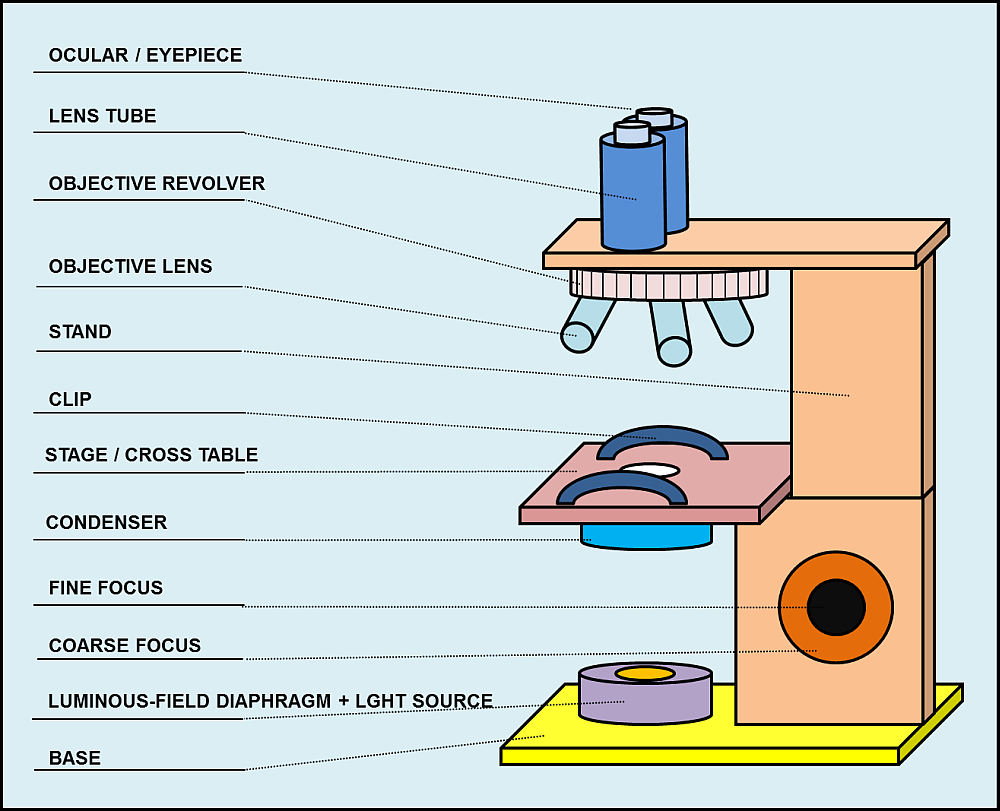
Picture: parts of a compound microscope
Ocular / Eyepiece
An eyepiece is that part of an optical system, which is directed to the viewer. It is a construction of at least one or more lenses. The function of the eyepiece in a microscope is to convert the real- enlarged-intermediate-image from the objective into an enlarged-virtual-image. You can find more details to this procedure in the chapter: “Enlargement in a Microscope”.
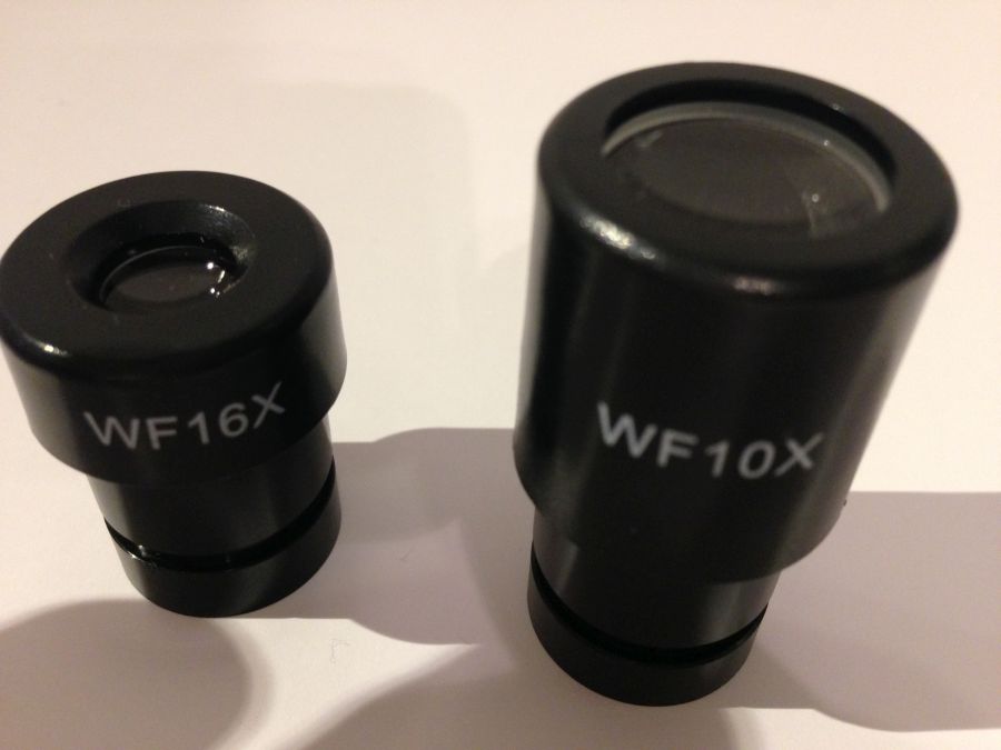
The size of the outpassing cone of light is adjusted to the size of the human eye. In an ideal case, the exit pupil is not larger, so that the complete pencil of rays can enter the eye.
Lens tube
The lens tube is connected with the eyepiece and it´s main task is to hold it.
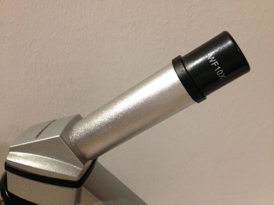
Microscopes normally operate with a lens tube length of 160 millimeters. Newer and especially professional microscopes use a longer lens tube. Thus, one can easily insert additional components like color filters or polarizers between eyepiece and objective.
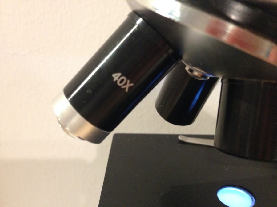
Objective revolver
Objective revolvers are used in microscopes with multiple objective lenses, that have different magnification factors.
By spinning the revolver, one can chose the lens with the desired enlargement level.
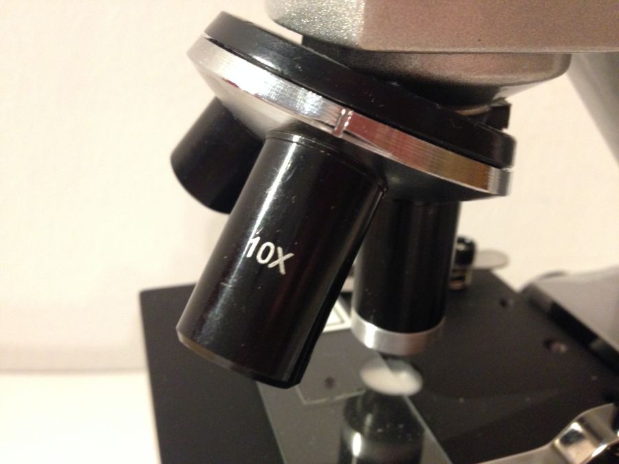
Objective lens
An objective (lens) is that part of an optical system, which is directed to the object. It´s task is to collect the ligh rays, that are reflected from the observed item. The objective generates a real-optical image.
Stand
The stand is connected with all the components and is holding them together.
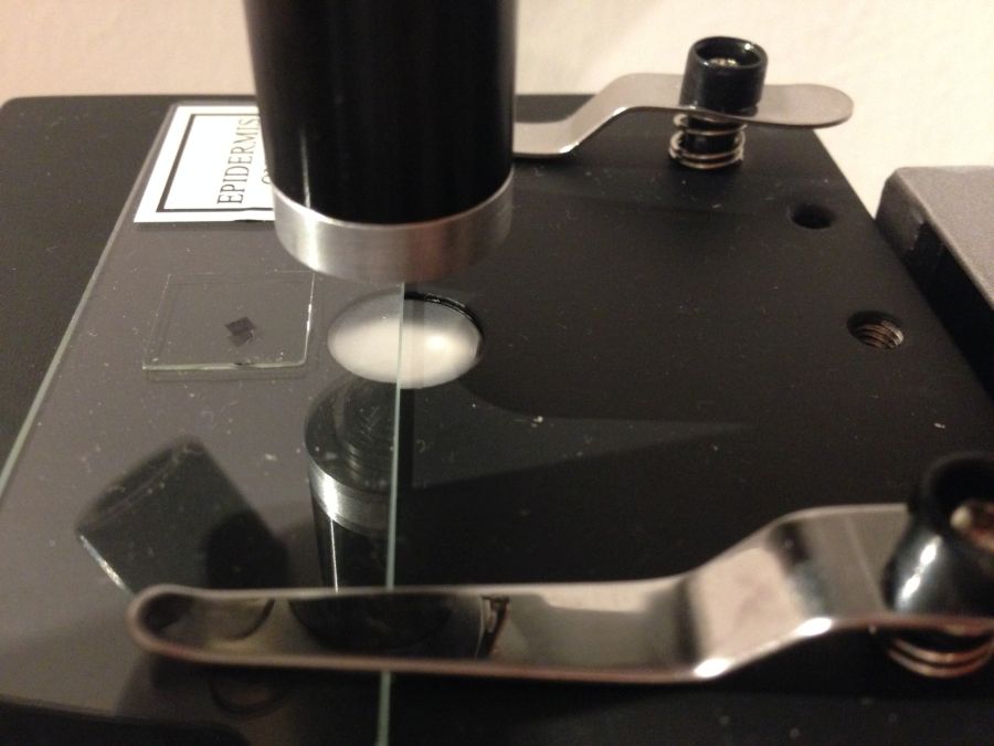
Clip
The clip serves as a holder for the object plate and makes sure, that it doesn´t get out of its place unintentionally.
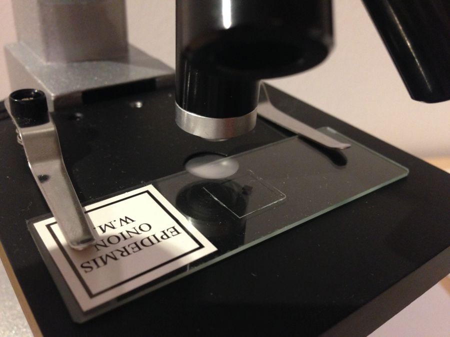
Microscope stage / cross table
On the stage, one can place the object plate with the cover glass on it. By shifting the plate, one can chose the part of the object, which one wants to look at.
Microscopes of higher quality sometimes use a cross table as stage. This allows to shift the object plate via adjusting screw, instead of doing it by hand. “Cross table” is technical term. It is a construction, where a table is installed into a system of rails. There are adjusting screws, which can be used to move the table very precisely. The screws are provided with a measuring scale, so one can always find again a certain point of the object.
Condenser
The condenser bundles the rays from the light source, so they are projected equally on the object. Thuse, every part of the object is illuminated on the same brightness level.
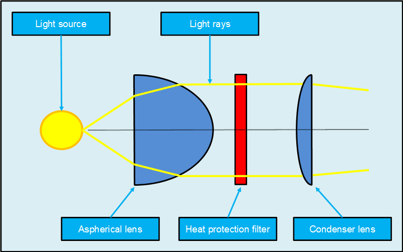
Condensers normally consist of one or two lenses. These lenses fractionize the light and all the rays leave as parallel beams. An aspherical lens makes sure, that no aberrations occur. This guarantees a better image quality. Their production costs are higher than those of normal lenses. They can be another criteria that differentiates high-performance microscopes from cheap microscopes.
Another way of to perfectly reflect light are optical filters with dielectric surface. Such Bragg mirrors consist of thin layers with sometimes high and sometimes low refractive index. They enable a very high quality reflection of light rays, which makes the image sharper later and reduces lens errors prohyilactically.
Fine focus & Coarse focus
With the fine focus one can regulate the distance between object and objective, to achieve the necessary sharpness. The fine focus moves the stage only minimally – like the name already says.
Like the fine focus, the coarse focus also moves the stage to regulate the difference between object and objective. His task is, to catch the right distance roughly and quickly. The optimal sharpness can be adjusted with the fine coarse afterwards.
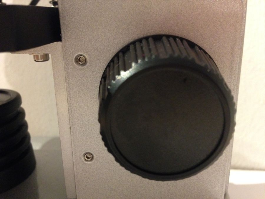
Luminous-field diaphragm
The luminous-field diaphragm can adjust the diameter of the light ray from the light source. This can prevent the object from being outshined.
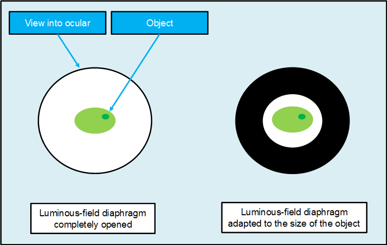
This can happen, when the object´s diameter is smaller than that of the viewing range.
Light Source
The early microscopes used concave mirrors to refrect light on the objects. Later, they used light bulbs. Most microscopes operate wiht LED ligth. The light source´s task is to illuminate the object evenly.

Base
The base guarantees the necessary stability to the microscope.
NEW!!! Free software: learn the stucture of the microscope
https://youtu.be/Zg4r-HcNnMM